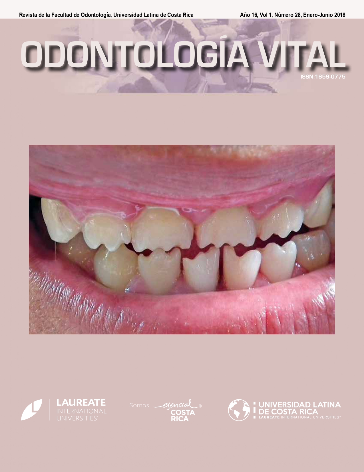
Diagnostic accuracy of two digital systems in the detection of carious lesions
DOI:
https://doi.org/10.59334/ROV.v1i28.160Keywords:
Dental Caries, digital radiography, sensitivity, specificityAbstract
Objective: To determine the accuracy in the diagnosis of interproximal and occlusal carious lesions of two digital systems: Charged Coupled Device (CCD) and Photo Stimulable Phosphor (PSP), using as a gold standard the histological evaluation.
Methods: 207 surfaces of teeth were evaluated using two digital systems: CCD (Planmeca ProSensor® HD) and PSP (VistaScan mini Easy Dürr Dental). The actual depth of the carious lesion was determined by the histological evaluation, using the Dinolite Microscope. To determine the accuracy, sensitivity, specificity, positive predictive value and negative predictive value were performed.
Results: The histological evaluation found 62 dental pieces with carious lesion on the occlusal surface, 38 pieces on mesial and 33 pieces on distal. The sensitivity in the occlusal surface was 95.15% for both systems, in mesial was 78.95% for CCD and 63.16% for PSP, in the distal surface was 75.76% for CCD and 78.79% PSP. The specificity for the surfaces evaluated with both systems was between 90-100%.
Conclusion: The diagnostic accuracy of the CCD and PSP digital systems were similar for the detection of occlusal and interproximal carious lesions. It is concluded that the modality of the image is not the factor that alters the diagnosis.
Downloads
References
Oropeza A, Molina N, Castañeda E, Zaragoza Y, Cruz D., (2012) Caries dental en primeros molares permanentes de escolares de la delegación Tiahuanaco. Rev. ADM. ; 69(3):63-8.
Rubio E, Cueto M, Suárez R, Frieyro J., (2006) Técnicas de diagnóstico de la caries dental. Descripción, indicaciones y valoración en su rendimiento. Bol Pediatr.; 46(1):23-31.
Hoyos M, Esperella A, Saavedra C, Espinoza H., (2013) Radiología de la caries dental. Rev. Actualización Clínica.; 38(1): 1857-62.
Ferjerskov O. (1997) Concepts of dental and their consequence for understanding the disease. Dent Oral Epidem.; 25(1): 5-12. https://doi.org/10.1111/j.1600-0528.1997.tb00894.x
Cuadrado D, Peña R, Gómez J. (2013) El concepto de caries: hacia un tratamiento no invasivo. Rev. ADM. ; 70(2):54-60.
Pontual A, Melo D, Almeida A, Bo Sciki F, Neto F. (2010) Comparison of digital systems and conventional dental film for the detection of approximal enamel caries. Dentomaxillofacial Radiology.; 39:431-6. https://doi.org/10.1259/dmfr/94985823
Veitía L, Acevedo A, Sánchez F. (2011) Métodos convencionales y no convencionales para la detección de lesión inicial de caries. Revisión Bibliográfica. Acta. Odontol. Venez.; 49(2):1-14.
Syriopoulos K, Sanderink G, Velder X, Van de Stelt P. (2000) Radiographic detection of approximal caries: a comparison of dental films and digital imaging systems. Dentomaxillofacial Radiology.; 29(1); 312-8. https://doi.org/10.1038/sj/dmfr/4600553
Abesi F, Mirshekar A, Moudi E, Seyedmajidi M, Haghanifar S, Haghighat N, et ál. (2012) Diagnostic accuracy of digital and conventional tadiography in the detection of non-cavitated approximal dental caries. Iran J Radiol.; 9(1):17-21. https://doi.org/10.5812/iranjradiol.6747
Saadettin K, Omer S, Senem S, Gamze C. (2011) An in vitro comparison of diagnostic abilities of conventional radiography, storage phosphor, and cone beam computed tomography to determine occlusal and approximal caries. Eur. J Radiol.;80(2): 478-82. https://doi.org/10.1016/j.ejrad.2010.09.011
Kamburoglu K, Senel B, Yuksel S, Ozen T. (2010) A comparison of the diagnostic accuracy of in vivo and in vitro photostimulable phosphor plate digital images in the detection of occlusal caries lesions. Dentomaxillofacial Radiology.; 39(1):17-22. https://doi.org/10.1259/dmfr/91657756
Mepparambath R, Bhat S, Hegde S, Anjana G, Sunil M, Mathew S. (2014) Comparison of proximal caries detection in primary teeth between laser fluorescence and bitewing radiography: An in vivo study. IJCPD. Dec; 7(3):163-7. https://doi.org/10.5005/jp-journals-10005-1257
Zhang Z, Qu X, Li G, Zhang Z, Ma X. (2011) The detection accuracies for proximal caries by cone-beam computerized tomography, film, and phosphor plates. Oral and Maxillofacial Radiology. Jan; 111(1):103-8. https://doi.org/10.1016/j.tripleo.2010.06.025
Cheng J, Zhang Z, Wang (2012) Detection accuracy of proximal caries by phosphor plate and cone-beam computerized tomography images scanned with different resolutions. Clinical Oral Invest.; 16(1):1015-21. https://doi.org/10.1007/s00784-011-0599-7
Sogur E, Baksi B, Mert A. (2012) The effect of delayed scanning of storage phosphor plates on occlusal caries detec-tion. Dentomaxillofacial Radiology.; 41(1):309-15. https://doi.org/10.1259/dmfr/12935491
Tarim E, Kucukyilmaz E, Ertas H, Savas S, Yircali M. (2014) A comparative study of different radiographic methods for detecting occlusal caries lesions. Caries Res.; 48(1):566-74. https://doi.org/10.1159/000357596
Kamburoglu K, Kurt H, Kolsuz E, Oztas B, Tatar I, Hamdi H. (2011) Occlusal caries depth measurements obtained by five different imaging modalities.; 24(1):804-13. https://doi.org/10.1007/s10278-010-9355-9
Abou L, Benedicto J, Luiz P. (2006) Evaluation of the effectiveness of clinical and radiographic analysis for the diagnosis of proximal caries for different clinical experience levels: comparing lesion depth through histological analysis. Braz J Oral Sci.; 5(17):1012-7.
Kamburoglu K, Tsesis I, Kfir A, Kaffe I. (2008) Diagnosis of artificially induced external root resorption using conventional intraoral film radiography, CCD, and PSP: an ex vivo study. RSS. ; 1(1):1-4.
Wiesi M, Hintze H , Wenzel A. (2007) Comparison of diagnostic accuracy of film and digital tomograms for assessment of morphological changes in the TMJ. Dentomaxillofacial Radiology., 36(1):12-17. https://doi.org/10.1259/dmfr/78486936
Saquete Martins P.R., Filho, Ferreira Da Silva L.C., Rabello Piva M., Machado Reinheimer D., Sue Dejean K. (sf) Spread of odontogenic infection originating from endoperio lesion. Endoperiodontal lesion – Case report. https://www.sigaa.ufs.br
Raja Sunitha V, Pamela Emmadi,[...],and Vijayalakshmi Rajaraman. (2008).The periodontal – endodontic continuum: A review. https://doi.org/10.4103/0972-0707.44046
Samar Abdul Hamed Bds, Msc. (2011). Repair of root canal perforation by different materials.
Syed Wall Peeran, Madhumania Thiruneervannan, Khaled Awidat Abdalla, Marei Hamed Mugrabi. (2013). Endoperio lesions.
Perdomo Masilly X., Ortiz Moncada C., Odalmis La O Salas N., Corona Carpio M.H., León Betancourt E.C. (2006). Principales aspectos clínicos de las afecciones encoperiodontales.
Downloads
Published
Issue
Section
License
Copyright (c) 2018 Milagros Montejo-Quirós, Andrés Agurto-Huerta

This work is licensed under a Creative Commons Attribution 4.0 International License.
Authors who publish with Odontología Vital agree to the following terms:
- Authors retain the copyright and grant Universidad Latina de Costa Rica the right of first publication, with the work simultaneously licensed under a Creative Commons Attribution 4.0 International license (CC BY 4.0) that allows others to share the work with an acknowledgement of the work's authorship and initial publication in this journal.
- Authors are able to enter into separate, additional contractual arrangements for the non-exclusive distribution of the Odontología Vital's published version of the work (e.g., post it to an institutional repository or publish it in a book), with an acknowledgement of its initial publication.
- Authors are permitted and encouraged to post their work online (e.g., in institutional repositories or on their website) prior to and during the submission process, as it can lead to productive exchanges, as well as earlier and greater citation of published work.


