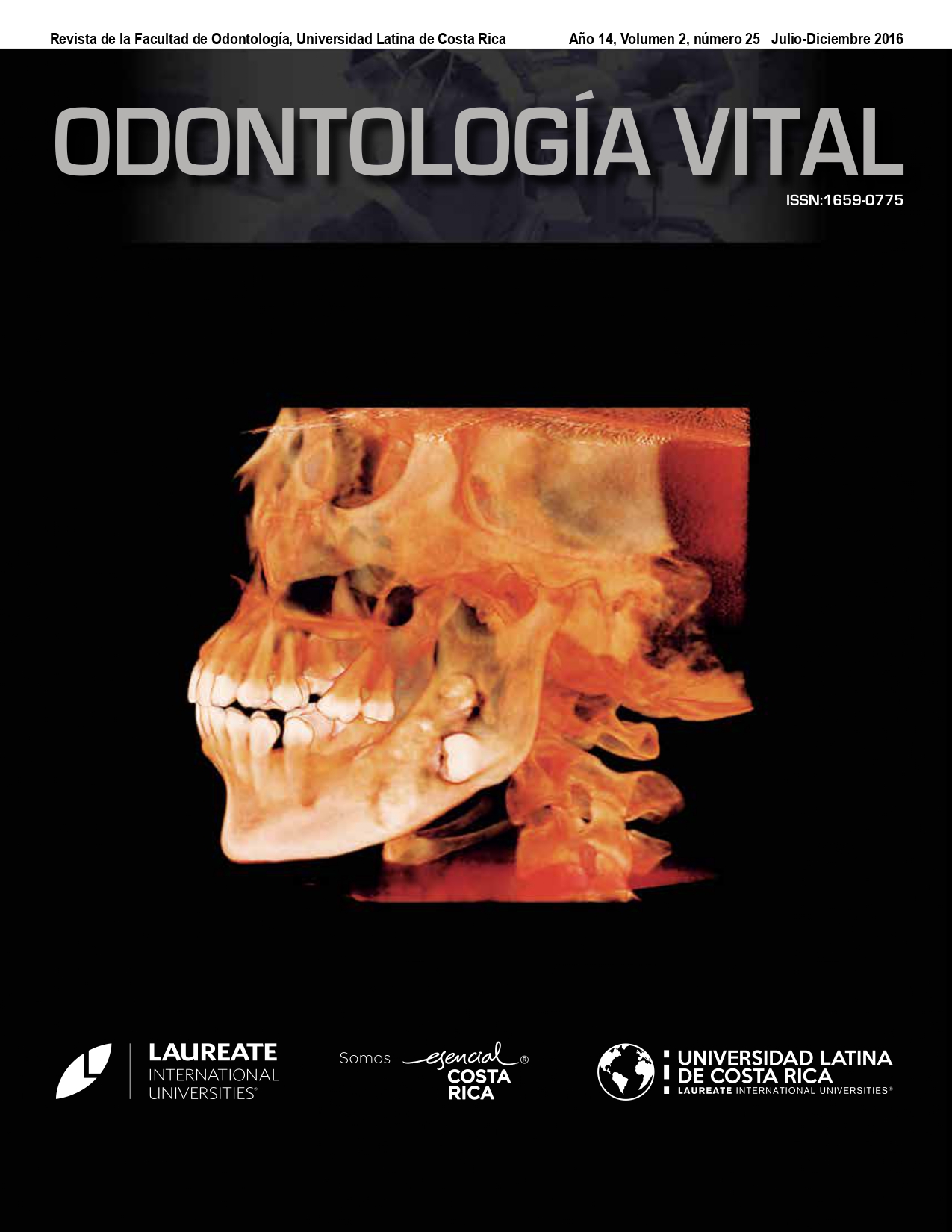
Mucocele del seno maxilar: Reporte de caso
DOI:
https://doi.org/10.59334/ROV.v2i25.243Palabras clave:
Mucocele, seno maxilar, Caldwell LucResumen
Los mucoceles maxilares son lesiones que tienen un crecimiento gradual. Son de tipo quístico y contienen secreciones mucoides que causan erosiones a estructuras vecinas al aumentar su tamaño. Aunque la mayoría de las veces son completamente asintomáticas, pueden presentarse síntomas como dolor facial opaco, inflamación en la mejilla, y obstrucción nasal. Estos síntomas y cambios en la simetría facial se hacen presentes cuando hay erosión significativa de estructuras anatómicas circundantes. EL seno maxilar es el sitio menos frecuente donde se forman , y su diagnóstico se realiza con la ayuda de una tomografía computadorizada. La remoción quirúrgica completa es el mejor tratamiento indicado.
Descargas
Referencias
Álvarez RJ., Liu NJ, Isaacson G. (1997). Pediatric ethmoidal in cystic fibrosis: long term follow up of reported case. Ear nose throat J.; 76(8):538-46. https://doi.org/10.1177/014556139707600810
Busaba NY, Kieff D. (2002). Endoscopic sinus surgery for inflammatory maxillary sinus disease. Laryngoscope , 112:1378–1383. https://doi.org/10.1097/00005537-200208000-00010
Busaba NY, Salman SD (1999). Maxillary sinus mucoceles: Clinical presentation and long-term results of endos-copic surgical treatment. Laryngoscope, 109:1446–1449. https://doi.org/10.1097/00005537-199909000-00017
Chen TM, Lee TJ, Huang TS. (1997). Endoscopic sinus surgery for the treatment of frontoethmoidal mucocele com-plicated with orbital abscess. Changgeng Yi Xue Za Zhi; 20: 39-43.
Dispenza C, Saraniti C, Caramanna C, Dispenza F. (2004). Endoscopic treatment of maxillary sinus mucocele. Acta Otorhinolaryngol Ital; 24: 292-96.
Fatma Caylakli , Haluk Yavuz (2006). Alper Can Cagici and Levent Naci Ozluoglu: Review Endoscopic sinus sur-gery for maxillary sinus mucoceles; Head & Face Medicine. 2:29. https://doi.org/10.1186/1746-160X-2-29
G Mohammadi, MD* M R Sayyah Meli, MD, M Naderpour, MD (2008). Endoscopic Surgical Treatment of Parana-sal Sinus Mucocele Med J Malaysia Vol 63 No 1 March
Har-el G (2001): Endoscopic management of 108 sinus mucoceles. Laryngoscope, 111:2131–2134. https://doi.org/10.1097/00005537-200112000-00009
Kim, YS; Kim, K; Lee, JG., Yoon JH, Kim CH. (2011). Paranasal sinus mucoceles with ophthalmologic manifesta-tions: a 17-year review of 96 cases. Am J Rhinol Allergy; 25:272–5. https://doi.org/10.2500/ajra.2011.25.3624
Lee TJ, Li SP, Fu CH, Huang CC, Chang PH, Chen YW, ChenCW. (2009). Extensive paranasal sinus mucoceles: a 15-year review of 82 cases. Am J Otolaryngol Head Neck Med Surg.; 30:234–8. https://doi.org/10.1016/j.amjoto.2008.06.006
Lucas Gomes Patrocinio, Priscila Garcia Damasceno, José Antonio Patrocinio. (2008). Maxillary Mucocele in a 4 year old infant: Rev Bras Otorrinolaringo; 74(3):479. https://doi.org/10.1016/S1808-8694(15)30590-5
Loo JL, Looi AL, Seah LL. (2009). Visual outcomes in patient with paranasal mucoceles. Ophthal Plast Reconstr Surg. ; 25:126–9. https://doi.org/10.1097/IOP.0b013e318198e78e
Marks SC, Latoni JD, Mathog RH (1997): Mucoceles of the maxillary sinus. Otolaryngology Head and Neck Sur-gery. 117:18–21. https://doi.org/10.1016/S0194-59989770200-6
Serrano E, Klossek JM, Percodani J, Yardeni E, Dufour X. (2004). Surgical management of paranasal sinus muco-celes: a long-term study of 60 cases. Otolaryngol Head Neck surg; 131: 133-40. https://doi.org/10.1016/j.otohns.2004.02.014
Sheth HG, Goel R. (2007). Diplopia due to maxillary sinus mucocele. Int Ophthalmol; 27:365-367. https://doi.org/10.1007/s10792-007-9082-5
Tseng C, Ho CY, Kao SH. (2005). Ophthalmic manifestations of paranasal sinus mucoceles. China MED Assoc June; 68: 260-64. https://doi.org/10.1016/S1726-4901(09)70147-9
Weber AL. (1998). Inflammatory disease of the paranasal sinuses and mucoceles. Otolaryngol Clin North Am ; 21: 421-37. https://doi.org/10.1016/S0030-6665(20)31513-9
Ying-Nan Chang and Bor-Hwang Kang (2010): Idiopathic Maxillary Sinus Mucocele; J Med Sci;30(1):033-035
Descargas
Publicado
Número
Sección
Licencia
Derechos de autor 2016 Roselena Elena Del Valle Granados, Efraín Cima García, Sergio Castro Mora

Esta obra está bajo una licencia internacional Creative Commons Atribución 4.0.
Los autores que publican con Odontologia Vital aceptan los siguientes términos:
- Los autores conservan los derechos de autor sobre la obra y otorgan a la Universidad Latina de Costa Rica el derecho a la primera publicación, con la obra reigstrada bajo la licencia Creative Commons de Atribución/Reconocimiento 4.0 Internacional, que permite a terceros utilizar lo publicado siempre que mencionen la autoría del trabajo y a la primera publicación en esta revista.
- Los autores pueden llegar a acuerdos contractuales adicionales por separado para la distribución no exclusiva de la versión publicada del trabajo de Odontología Vital (por ejemplo, publicarlo en un repositorio institucional o publicarlo en un libro), con un reconocimiento de su publicación inicial en Odontología Vital.
- Se permite y recomienda a los autores/as a compartir su trabajo en línea (por ejemplo: en repositorios institucionales o páginas web personales) antes y durante el proceso de envío del manuscrito, ya que puede conducir a intercambios productivos, a una mayor y más rápida citación del trabajo publicado.


