Fluoride. Antivet vs Hydrochloric Acid.
Differences between antivet and hydrochloric acid
DOI:
https://doi.org/10.59334/ROV.v1i37.473Keywords:
toxicity , endemic, emaciation, hydrochloric acidAbstract
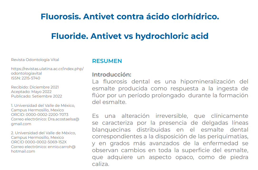
Introduction: Dental fluorosis is a hypomineralization of the enamel produced as a response to the ingestion of fluoride for a prolonged period during the formation of enamel. It is an irreversible alteration, which is clinically characterized by the presence of thin whitish lines distributed in the dental enamel corresponding to the arrangement of the perikymatias, and in more advanced stages of the disease, changes are observed throughout the surface of the enamel, which acquires an opaque, limestone-like appearance. In the most severe levels of dental fluorosis, the presence of hypomineralization, and the increase in the porosity of the dental enamel favors the loss of important portions of its structure, producing fractures, thus deteriorating the appearance and functionality of the affected teeth. The WHO recommends that the reference value for fluoride in drinking water is 1.5 mg / l. Fluorine is a halogen gas, the most electronegative of the elements in the periodic table, with atomic number 19. It practically does not exist free in nature, but is associated with other elements such as calcium and sodium. The main way that fluorine is incorporated into the human body is through the digestive tract. It is rapidly absorbed in the mucosa of the small intestine and the stomach, by a simple diffusion phenomenon. Once absorbed, fluorine passes into the blood and is distributed in the tissues, depositing preferably in hard tissues; it is eliminated through all excretion routes, mainly through urine.
Objective: to know how to differentiate the types of materials and to know the different methods for eliminating fluorine, as well as to show the difference between treatments.
Methodology: The type of study is explanatory and with which it is hoped to contribute to the development of scientific knowledge. Its realization supposes the desire to contribute to the development of scientific knowledge. It consisted of selecting 16 patients, male and female and of different ages between 15 and 40 years. They were randomly divided into 2 groups of 8 people each to be treated with 2 different products. The first group was treated with 18% hydrochloric acid and the second group with the Antivet brand.
Result and conclusion: Dental fluorosis is caused by excessive fluoride intake. The use of hydrochloric acid is corrosive, its aroma is penetrating and the patient's care is greater, since misuse when in contact with skin or mucosa will create necrosis. Antivet has disadvantages in cost and availability, but its advantage is that it provides greater safety in its handling.
Downloads
References
Elsa María Acosta Enriquez y Ennio Hector Carro Hernandez. (Junio 2021). Fluorosis. Antivet contra Ácido Clorhídrico. Odontología Vital, 1, 7. https://doi.org/10.59334/ROV.v1i37.473
Narro Robles, J., Meljem Moctezuma, J., Kuri Morales, P., Velasco González, M. G., González Roldán, J. F., Ruiz Matus, C., Mancha Moctezuma, C., Jiménez Corona, M. E., & Díaz Quiñonez, J. A. (2015). Patologías Bucales (SIVEPAB) 2015 - gob.mx. Retrieved April 23, 2022,
Salud Mexicana, S. de. (2015). Norma Oficial mexicana NOM-013-SSA2-2015. - amic dental. Para la prevención y control de enfermedades bucales. NORMA013. Retrieved April 23, 2022, Manual para el uso de fluoruros dentales en la República Mexicana. Secretaría de Salud. 2006.
Ham CC, Hart TC, Dupont BR, Chen JJ, Sun X, Quian Q. Moning human enamelin DND, chromosomal localization and analysis of expression during tooth development. J Dent Ress 2000; 73 (4): 912-9. https://doi.org/10.1177/00220345000790040501
Finchom AG, Simmer JP. Amelogenin, proteins of developing dental enamel. Ciba Found Symp 2000; 205: 118-30. https://doi.org/10.1002/9780470515303.ch9
Dean H. Classification of mottled enamel diagnosis. J Am Dent Assoc. 1934; 21:1421-1426. https://doi.org/10.14219/jada.archive.1934.0220
Young MA. Dental health education-whither? J Am Dent Assoc. 1963 Jun; 66:821-4
(Deán 1934). Colaborando en Jackson 1975) Young (1963).
Salgam AM, Ozbaran HM, Salgam AA.A comparación of mesio distal Crown dimensions of the permanent teeth in subjects with and without. Fluorosis. Eur J Orthod: 2004 JUN; 26(3): 279-81. https://doi.org/10.1093/ejo/26.3.279
Espinosa R. Análisis químico del esmalte fluorótico. Odontologia Actual. 194b; Año ll N° 9:7-11
Diamond M. Anatomía Dental. Ed.2.Mex:Uthea.1962 Eager J. Denti di Chiaie teeth (Chiaie teeth). US Pub Health. 1901.Rep 16:2576
Cutress TW, Suckling GW. Differential diagnosis of dental fluorosis. J Dent Res [Internet]. 1990;69:714–720; discussion 721. https://doi.org/10.1177/00220345900690S138
Norma Oficial Mexicana para la prevención y control de enfermedades bucales. Nom 013-ssa2-21 de enero 1994- 1999 Largent EJ (1961). Fluorosis the health aspects of fluorine compounds Columbus Ohio State Univ. Press. Logan WHEy Kronfeld R. Chronology of the human dentition, J.A.D.A. 1933; 20:379
Yoon SH, BrudevolD F, Gardner DE y Smith FA. Distribution of fluoride in teeth from áreas with different levels of fluoride in the water supply. J Dent Res. 1960;39:845-56. https://doi.org/10.1177/00220345600390041101
Aoba T. Fejerskov O. Dental fluorosis: chemistry and biology: Crit Rev Oral Biol Med. 2002;13 (2): 155-70. https://doi.org/10.1177/154411130201300206
Osborn JM, Tencate AR. Dentine sensitivity. En: Advances dental histology. 4ed. Bristol: Editorial Wright PSG; 2003.p. 109-17. (Sat Chell PG y Cols 2002).
Fejerskov O, Thylstrup A, Larsen M. Clinical and structural features and possible pathogenic mechanisms of dental fluorosis. Scand J Dent Res. 1977;855:22-30. https://doi.org/10.1111/j.1600-0722.1977.tb02110.x
Rugh A. Impact of orthognathic surgery on normal and abnormal personality dimensions: A “ years follow up study of 61 patients Am: J. Orthod. Dentofac. Orthop: 1990;98:313-222. https://doi.org/10.1016/S0889-5406(05)81488-X
Mclnnes J. Removing Brown stain from teeth: Ariz Dent. J. 1966 May 15;12(4): 13-5
Espinosa R. TratamIento de la fluorosis dental y su relación con los diferentes grados. Odontologia Actual. 1995b; Año ll, N°8:7-15
Espinosa R. TratamIento de la fluorosis dental y su relación con los diferentes grados. Odontologia Actual. 1995b; Año ii, N°8:7-15
Espinosa R. Esmalte fluorótico: Análisis al microscopio electrónico. Odontologia Actual. 1995d Sep-Oct;7-12
Salgam AM, Ozbaran HM, Salgam AA.A comparación of mesio distal Crown dimensions of the permanent teeth in subjects with and without. Fluorosis. Eur J Orthod: 2004 JUN; 26(3): 279-81. 25. https://doi.org/10.1093/ejo/26.3.279

Downloads
Published
License
Copyright (c) 2022 Elsa Acosta Enriquez, Ennio Carro Hernández

This work is licensed under a Creative Commons Attribution 4.0 International License.
Authors who publish with Odontología Vital agree to the following terms:
- Authors retain the copyright and grant Universidad Latina de Costa Rica the right of first publication, with the work simultaneously licensed under a Creative Commons Attribution 4.0 International license (CC BY 4.0) that allows others to share the work with an acknowledgement of the work's authorship and initial publication in this journal.
- Authors are able to enter into separate, additional contractual arrangements for the non-exclusive distribution of the Odontología Vital's published version of the work (e.g., post it to an institutional repository or publish it in a book), with an acknowledgement of its initial publication.
- Authors are permitted and encouraged to post their work online (e.g., in institutional repositories or on their website) prior to and during the submission process, as it can lead to productive exchanges, as well as earlier and greater citation of published work.


